|
The heart is an extremely efficient pump that is electrically activated by a specialized group of cells (sinus node) capable of spontaneous excitation at periodic intervals. However, in certain situations, this normal activity can be hampered by arrhythmias, e.g., ventricular tachycardia (VT), during which the heart can beat as rapidly as 300 beats per minute. The most common mechanism of VT is the formation of a closed path of excitation feedback (reentry) around an existing inexcitable obstacle, e.g., a region of scar tissue.
We look at biologically realistic computational models of heart tissue to understand how low-amplitude electrical stimulation (pacing) can rapidly terminate VT. For ease of theoretical analysis and numerical computations, we focus on reentry in a one-dimensional ring of cardiac cells which is essentially the region immediately surrounding an anatomical obstacle.
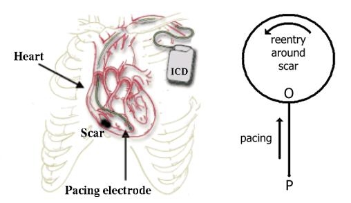 |
| Fig. 1. (Left) Schematic diagram of anti-tachycardia pacing in the heart using an ICD (adapted from a figure courtesy of Guidant Corp.). Note the non-conducting scar tissue (in black) occupying a significant portion of the ventricle. Pacing is usually applied via an electrode placed at the ventricular apex (the lowermost point of the ventricle in the figure). (Right) A simplified ring-and-sidebranch model of pacing. Reentrant activity occurring around a scar tissue is simplified into a wave going around a ring. The sidebranch joining the ring at O represents the external stimulation arriving from the pacing electrode located at P. |
Based on extensive computer simulations, we arrive at the conclusion that successful termination of VT requires the pacing to induce transient inhomogeneities in the reentry circuit. Generating such dynamical inhomogeneities (e.g., a zone of slow propagation) can be controlled by the pacing parameters. E.g., the number of pacing pulses has an optimal value, as using larger number of pulses often cause further conduction blocks restarting the reentry. The pacing frequency has to be carefully chosen also: it has to be higher than the VT frequency but it cannot be too high, as the propagation of high frequency waves causes instability and wave breakup, leading to new arrhythmias through formation of spiral waves.
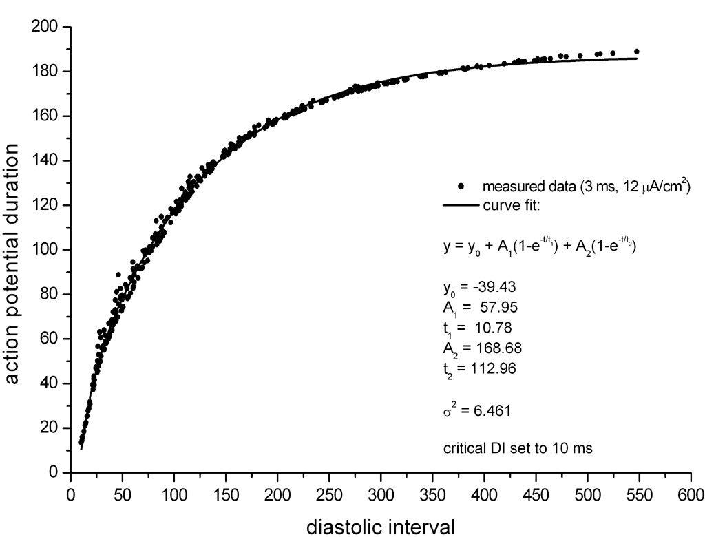 |
Fig. 2. The restitution curve obtained from a model of heart tissue. |
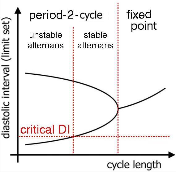 |
Fig. 3.Bifurcation diagram. |
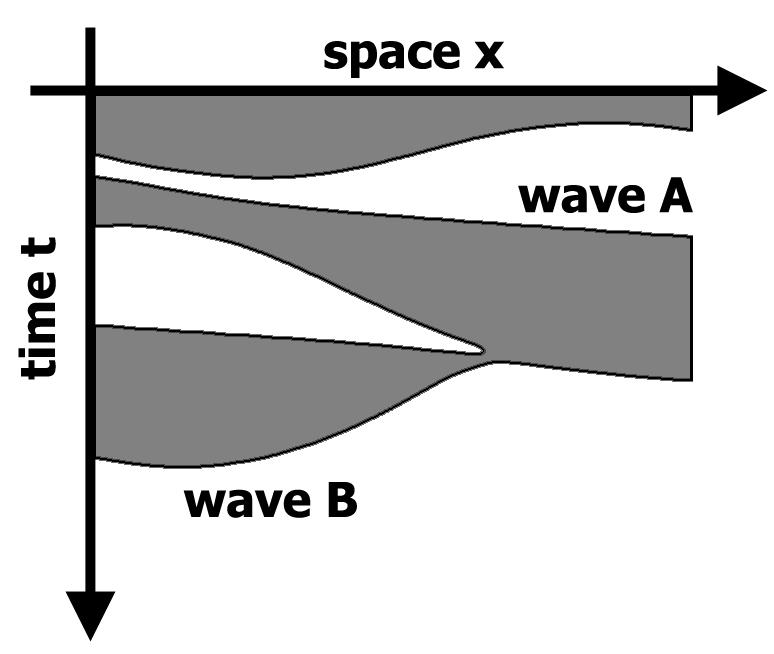 |
Fig. 3.Conduction block and hence, successful terminaition of reentry. |
The ultimate goal of anti-tachycardia pacing is to terminate reentry with pulses of smallest magnitude in the shortest possible time with the lowest probability of giving rise to faster arrhythmias. The constant frequency pacing investigated uptill now is only a partial solution to this end, and a more efficient algorithm might have to adjust the pacing intervals on a beat-to-beat basis. The results of our investigation is aimed towards answering how such an optimized pacing scheme maybe designed.
Publications: